 In 2020, the worldwide prevalence of glaucoma was estimated to be about 79.6 million people, yet many people do not understand this condition or how to identify the signs.
Glaucoma is an eye disease that affects the optic nerve. It most commonly occurs when pressure inside the eye is too high due to a backup of fluid, but it can also result from an injury or infection in the eye. Glaucoma can cause irreversible vision loss and blindness when intraocular pressure or IOP (pressure in the eye) builds up and damages the nerve in the back of the eye called the optic nerve. It is the optic nerve that sends visual information from your eye to your brain which is vital for good vision.
During the early stages of this disease, patients rarely ever have symptoms even as damage is occurring to the optic nerve, and this is why it is known as the silent thief of sight. As the disease progresses, vision loss starts with loss of the peripheral, or side vision, with patients reporting having some difficulty seeing things on one or both sides and if left untreated, leads to complete vision loss.
Buildup of Eye Pressure
There is a clear liquid inside the front part of the eye called aqueous humor which functions to provide nutrients to the eye as well as clear out waste. As new aqueous humor is produced and flows into your eye, the same amount should drain out through the trabecular meshwork (tiny canals that drain fluid from the eye). The area where the fluid drains out of the eye is called the drainage angle and is where the iris (coloured part of the eye) meets the cornea (transparent covering that lets light into the eye). This process of aqueous humor production and drainage keeps the intraocular pressure stable and keeps the eyes healthy and functioning normally. If there is an imbalance to this process, fluid can build up leading to increased pressure inside the eye. If the pressure is not brought under control, the optic nerve can be damaged and your vision permanently impaired.
Glaucoma Defined
There are two main types of glaucoma, open-angle and angle-closure glaucoma. Open-angle glaucoma is the most common form of glaucoma and is caused by a blockage in the drainage site of the eye. In this type of glaucoma, the buildup of intraocular pressure is gradual, and most times is symptomless. Angle-closure glaucoma less common but is considered a medical emergency. This type of glaucoma is characterized by a bulging of the iris which partially or completely narrows the angle of drainage formed between the iris and cornea. Symptoms may occur suddenly or gradually such as severe pain in the eye or forehead, decreased vision or blurred vision, headache, nausea and vomiting.
Risk Factors
To avoid irreversible vision loss, regular eye checks are recommended especially for people with underlying risk factors for glaucoma which include:
- High Intraocular Pressure (IOP): Lowering eye pressure is the only modifiable risk factor of glaucoma.
- Race: People of African ancestry have a greater risk of having glaucoma, its onset is earlier and disease progression is faster and more resistant to treatment.
- Age: Prevalence increases with age.
- Family history: Having a first degree relative (parent, sibling, child) with glaucoma is a risk factor for development of glaucoma.
- Systemic diseases: People with diabetes have double the risk of developing glaucoma. Hypertension (high blood pressure) may also put patients at risk of glaucoma through the narrowing or blocking of small vessels, several of which are found in the eye.
- Central cornea thickness: A thinner cornea (transparent part of the eye that covers the iris and pupil) may indicate less rigid support structures around the optic nerve head leading to an increased risk of damage.
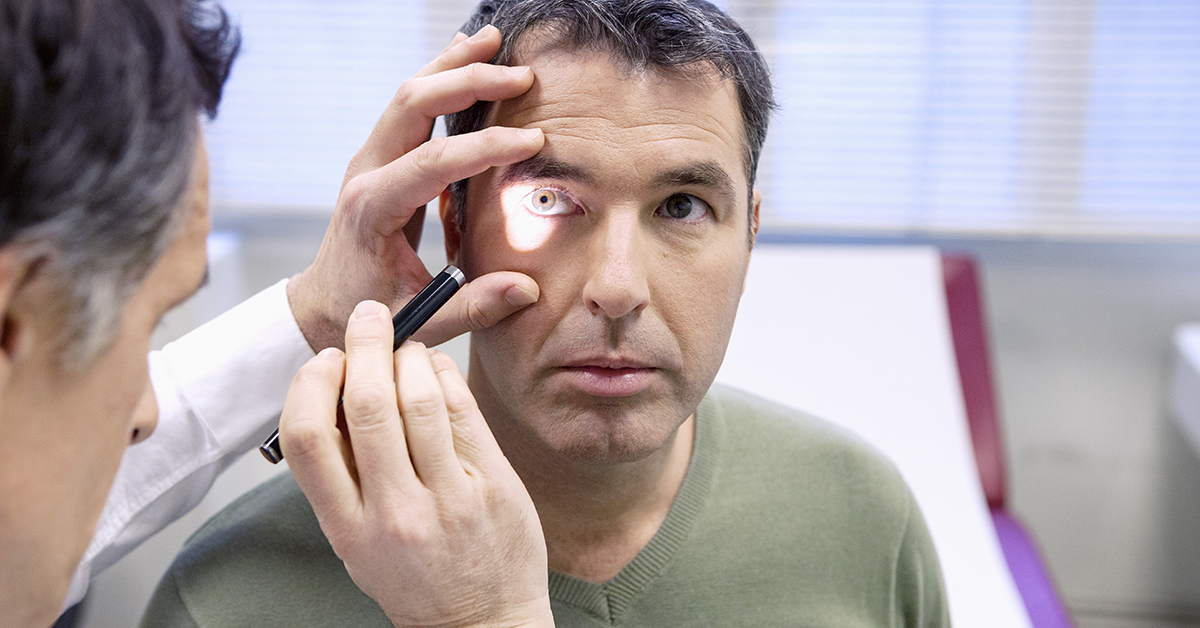
Eye Tests For Glaucoma
Several eye tests can be performed by an eye specialist to help diagnose glaucoma.
- Tonometry: a measurement of eye pressure.
- Visual Field Test: to tell if there is any loss to the patient’s field of vision from glaucoma and how much have been lost. It helps the doctor determine the rate of disease progression and to tailor the treatment to the stage of glaucoma.
- Glaucoma Imaging Test: a photograph of the optic nerve with a digital camera to map and document the health of the optic nerve.
- Cornea Thickness Test: a measurement of the central cornea thickness. Patients with abnormally thin central cornea have been found to have more damage to the optic nerve.
- Angle Test: a measurement of the angle where the iris meets the cornea leading to a diagnosis of “open-angle” glaucoma or “closed-angle” glaucoma.
Treatment
The primary goal of glaucoma treatment is to reduce the intraocular pressure (IOP) thereby reducing the progression to vision loss. Medicated eye drops are the treatment of choice and there are various types available depending on the patient’s causes of their increased eye pressure. Combination products are also available. If the eye drops are unsuccessful or not tolerated by the patient, laser or surgical procedures can be considered.
- Prostaglandin Analogues (Examples: latanoprost, bimatoprost, travoprost)
Increase aqueous humor drainage and are considered first-line treatment. - Beta-blockers (Examples: betaxolol, timolol)
Decrease aqueous humor production and are the second most effective class of medications after the prostaglandin analogues. - Alpha Adrenergic Agonists (Example: brimonidine)
Increase drainage and decrease production of aqueous humor. - Carbonic Anhydrase Inhibitors (Examples: brinzolamide, dorzolamide)
Decrease aqueous humor production. - Cholinergic Agonists (Example: pilocarpine):
Increase aqueous humor drainage.
Conclusion
While there is no cure for glaucoma, early treatment can stop optic nerve damage and protect a patient’s vision. For this reason, regular comprehensive eye exams are recommended for patients, especially those with risk factors of glaucoma so treatment can be started early. Even for patients who are not in the glaucoma high-risk group, regular comprehensive eye exams are recommended from the age of 40 as this can help to detect other age-related eye diseases that may occur, such as cataract, age-related macular degeneration and diabetic retinopathy. If you have questions about glaucoma or its treatment, speak to your doctor, eye care specialist, or pharmacist for more information.